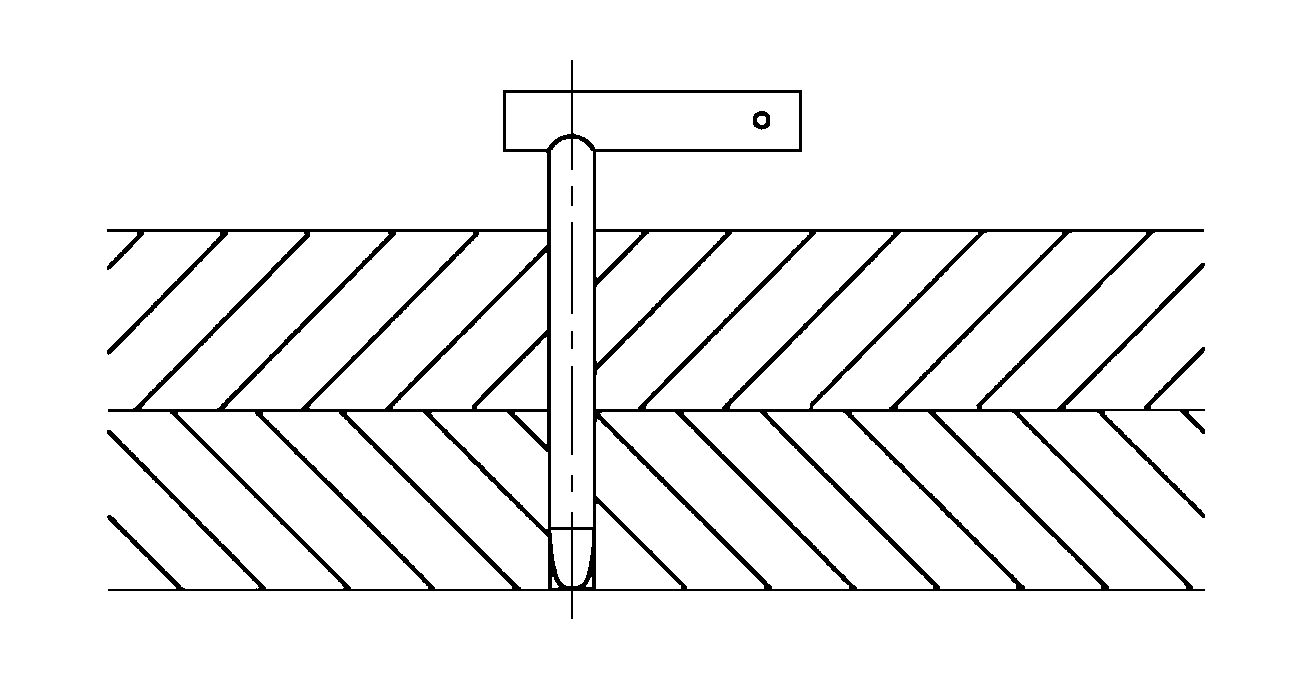
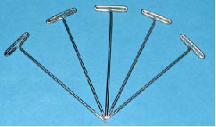
Left): Porcine ventricle sample, epicardium side up, mounted to
Download scientific diagram | (Left): Porcine ventricle sample, epicardium side up, mounted to the silicone lined fixture with Tpins. (Right): Porcine aorta sample, intima side up, mounted to the silicone lined fixture with T-pins. (Both): 0.25 in diameter steel ball upper member as test probe. from publication: PolyJet 3D Printing of Tissue Mimicking Materials: An Investigation of Characteristic Properties of 3D Printed Synthetic Tissue | Current anatomical 3D printing has been primarily used for education, training, and surgical planning purposes. This is largely due to the models being printed in materials which excel at replicating macro-level organic geometries; however, these materials have the drawback | 3D Printing, Tissue and Subcutaneous Tissue | ResearchGate, the professional network for scientists.

Anatomical and molecular mapping of the left and right ventricular
Pericardial Interventions: Pericardiocentesis, Balloon

The epicardial delivery of cardiosphere derived cells or their

Intact myocardial preparations reveal intrinsic transmural
/images/vimeo_thumbnails/258077871/4XOZEeUBEBJPMdBTvFzbcA_overlay.jpg)
Layers of the heart: Epicardium, myocardium, endocardium

Medicina, Free Full-Text
Frontiers Porcine Organotypic Epicardial Slice Protocol: A Tool
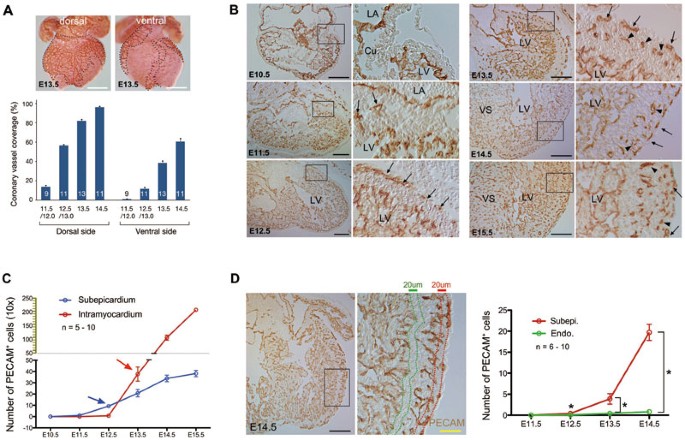
Subepicardial endothelial cells invade the embryonic ventricle

Heart Anatomy Anatomy and Physiology II

Section levels of the left ventricle.

Biventricular biaxial mechanical testing and constitutive

Preparation of a small mammalian (rat) left ventricular tissue

Neuroanatomy of the Pig Cardiac Ventricles. A Stereomicroscopic